Unveiling the Precision Attack: A Step-by-Step Guide to How Killer T Cells Destroy Cancer in 3D
Imagine a microscopic battlefield where a single cell must decide life or death. Scientists have now captured this moment in stunning 3D, revealing how the body’s killer T cells execute a perfectly choreographed strike against cancer. This guide will walk you through the hidden steps of that attack, from target recognition to final destruction—using the same molecular clues that researchers recently unveiled.
What You Need
- Basic biology grasp – Familiarity with cells, the immune system, and proteins helps, but curiosity is enough.
- Access to the research – The original 3D model (often published with open-access datasets) or a visual summary (e.g., from Nature or a university press release).
- Patience for scale – The process happens in seconds to minutes; understanding it requires slowing down our perspective to nanoscale time.
- Optional: 3D viewer software or animated video of the immunological synapse for a richer experience.
Step-by-Step Guide to the 3D Attack Mechanism
-
Step 1: Recognize the Target
A killer T cell patrols the body by scanning for fragments of foreign or abnormal proteins. When it encounters a cancer cell, the T cell’s surface receptor locks onto a specific antigen presented by the cancer cell’s MHC class I molecule. This initial binding is gentle but precise—like a key finding its lock. The 3D view shows that even this first contact is not random; the T cell reaches out with microvilli-like protrusions to sample the surface.
Source: www.sciencedaily.com -
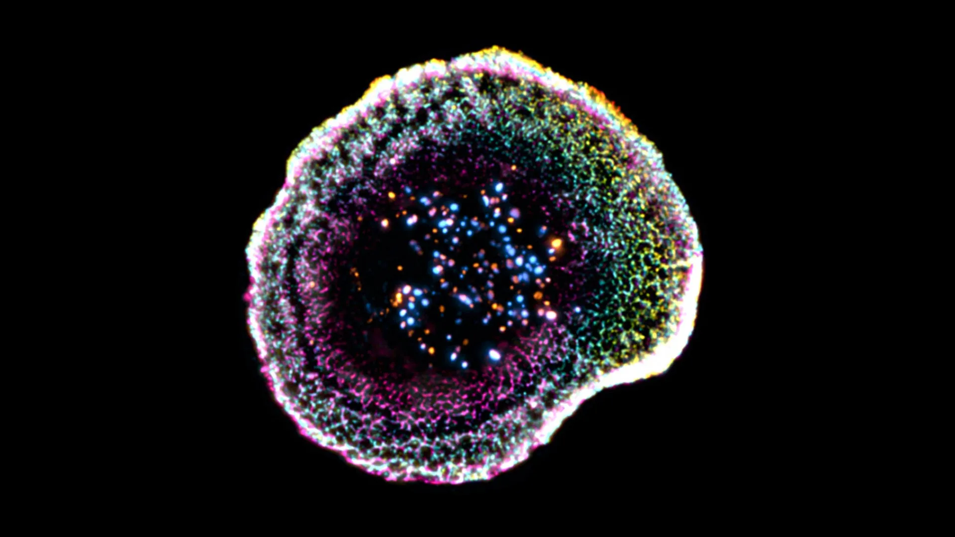
Step 2: Form the Immunological Synapse
Recognition triggers a dramatic reorganization. The T cell flattens against the cancer cell, forming a highly organized contact zone called the immunological synapse. In the new 3D images, researchers saw that this synapse is not a flat disk but a bullseye of concentric rings—each ring packed with different adhesion molecules and signaling proteins. This structure ensures the T cell stays tightly connected while avoiding damage to neighboring healthy cells.
-
Step 3: Polarize the Cytotoxic Granules
Inside the T cell, small membrane-bound bags called cytotoxic granules begin migrating toward the synapse. Powered by motor proteins along microtubules, they move like freight trains to a central hub. The 3D reconstruction captured this polarization in real-time, showing granules clustering directly under the contact zone—a crucial step that concentrates the killing machinery precisely where it is needed.
-
Step 4: Release Perforin and Granzymes
When enough granules have gathered, the T cell fuses them with its own membrane at the synapse, releasing two types of molecules. First, perforin forms pores in the cancer cell’s membrane, like punching holes in a balloon. Then granzymes (serine proteases) enter through those pores. The 3D view revealed that this release is not a chaotic spray but a directed stream, guided by the synaptic architecture to ensure not a drop of deadly enzyme leaks onto innocent bystanders.
-
Step 5: Trigger Apoptosis (Programmed Cell Death)
Once inside, granzymes activate a cascade of caspases—executioner proteins that slice up the cancer cell’s DNA and structural components. The cell begins to shrink, its membrane blebs outwards, and its nucleus fragments. This clean, programmed suicide avoids the inflammation that would occur if the cell simply burst. The 3D time-lapse shows the cancer cell collapsing inward, like a deflating balloon, while neighboring cells remain untouched.
-
Step 6: Detach and Recycle
After the kill, the T cell breaks contact, leaving behind the dead cancer cell for macrophages to clear. The T cell now has elevated cytotoxic granule levels ready for the next target. The 3D images even captured the recycling of membrane components at the synapse, showing how the T cell rapidly resets its attack machinery. This efficiency allows a single T cell to destroy multiple cancer cells in succession.
Tips and Insights from the 3D View
- Precision is key. The synapse’s bullseye pattern is not accidental—it prevents collateral damage. Think of it as a surgical laser versus a flamethrower.
- Watch the timing. Total attack can happen in under 10 minutes. But the 3D data shows that the most critical moments (steps 2–4) last only a few seconds.
- Implications for therapy. Understanding this choreography helps scientists design better immunotherapies, such as CAR-T cells that can be engineered to form even more efficient synapses.
- Learn from the model. If you have access to the original 3D dataset, explore it slice by slice. Notice how the T cell membrane is studded with tiny dimples and ridges that correspond to molecular clusters.
- Keep an eye on Step 1 and Step 4 – they are often the bottlenecks in artificial T cell designs.
By walking through these steps, you now see that a killer T cell’s attack is anything but random. It is a masterful dance of molecules—a war waged at the nanoscale with surgical precision, captured for the first time in three dimensions.
Related Articles
- How to Reduce Your Baby's Exposure to PFAS in Formula
- Mastering Log Noise Reduction with Adaptive Logs Drop Rules
- Preserving the American Dream: Challenges and Paths Forward
- Microscopic Gut Particles: Could They Hold the Key to Aging and Chronic Disease?
- Isomorphic Labs: Alphabet’s $2 Billion AI Bet to Revolutionize Drug Discovery
- FDA Faces Critical Talent Gap After Mass Firings, Agency Struggles to Rehire
- Disparities in Extreme Heat: Why Black Americans Face Greater Risks
- From the Track to the Screen: Apple's Strategic Play in Formula 1 Racing